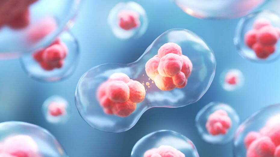
In modern IVF applications, genetic procedures have recently become quite popular and have contributed to pregnancy rates. As a result of the development of bio-technology, genetic embryo selection practices have been introduced to increase the success of IVF. PGT procedure is performed in 3 ways:
- With egg biopsy (polar body biopsy)
- PGT from the embryo of the clivage period
- From the embryo of the blastocyst period: Trophoectoderm Biopsy
What is PGT? How is it performed?
The embryo obtained during the IVF process contains a total of 8 cells called blastomeres when it reaches the 3rd day. This embryo is called the clivage period embryo. The blastomere taken from the clivage embryo is sent for genetic examination and the transfer process is performed or canceled according to the result. In the blastocyst period embryo, there is a trophoectoderm layer where the baby’s peer will develop in the later period. From this layer, a biopsy is taken with the help of a special syringe and sent for genetic examination. These procedures are performed separately for each embryo.
CGH Technique
With this technique, 3rd or 5th day embryos are examined with microarray-based comparative genomic hybridization technique and genetic analysis of all chromosomes is performed. After 24 hours, according to the results obtained, the healthy embryo is transferred to the mother’s womb. With this method, while live birth rates have increased, pregnancy losses have decreased.
What are the diseases that can be diagnosed with PGD?
- Single gene disorders.
- Cystic Fibrosis
- Alpha 1 antitrypsin deficiency,
- Muscular dystrophy
- Chromosomal Irregularities.
- Trisomies (embryos with a genetic structure consisting of 47 chromosomes), Triploidy (46 chromosomes are present in the normal person, 69 chromosomes in the trpiliodi), etc.
To whom is PGT recommended?
This procedure is a technique recommended for couples who are at high risk of having genetic problems in their baby. Thanks to this technique, embryos are genetically screened before being placed in the uterus. In this way, there is no need to wait for genetic analysis while the pregnancy continues, as in the past.
- Older age woman (with advancing age, the likelihood of genetic problems in the embryo and the risk of miscarriage increases)
- Balanced chromosomal translocations that do not cause phenotypic problems in the parents (may occur as a disease in the embryo)
- Recurrent IVF failures.
- Recurrent pregnancy losses.
- Unexplained Infertility.
- Severe male infertility.
- Y chromosome defect (deletion)
- Couples with abnormal embryos


